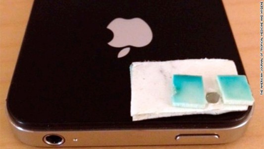
iPhone-ul a devenit din ce in ce mai popular in randul medicilor care folosesc diverse accesorii pentru a diagnostica boli, pentru a tine evidenta pacientilor, pentru a le oferi informatii suplimentare despre boli si pentru diverse alte functii. In Tanzania medicii sunt si mai inventivi, iar cu ajutorul unui iPhone, a unei lentile de camera, a unei benzi adezive si a unei lanterne au reusit sa confectioneze un microscop cu ajutorul caruia au verificat 200 de mostre ale unor elevi suspectati de a avea viermi intestinali.
In a discovery that provides doctors in remote areas with an alternative, scientists recently created a microscope using an iPhone, tape, flashlight and camera lens. They then used it to diagnose intestinal worms in about 200 students in Pemba Island, off Tanzania’s eastern coast. “To our knowledge, this is the first time the mobile phone microscope had been used in the field to diagnose intestinal parasitic infections,” said Isaac Bogoch, an internal medicine specialist at Toronto General Hospital.
iPhone 4S a fost utilizat pentru a verifica acele mostre, orice alt smartphone cu o camera decenta si functie de zoom ar putea face acelasi lucru, insa ca de obicei, terminalele Apple sunt la indemana pentru lucruri demne de atentia globului. Mai jos aveti explicat modul in care poate fi confectionat un microscop pentru smartphone-uri si, conform unui doctor canadian, aceasta ar fi prima oara cand un microscop fabricat pe loc este utilizat pentru o asemenea diagnosticare.
To convert the phone into a microscope, scientists put double-sided tape over the iPhone camera lens. They then pierced a hole in the tape and centered a tiny $9 lens over the phone’s camera lens. Using a flashlight for illumination, they held up stool samples against the lens using the double-sided tape to hold slides in place, and studied them for intestinal parasites through the phone’s screen. Bogoch said they diagnosed 70% of the infections when compared with the results of a conventional laboratory microscope.


